soon3

الاثنين، 30 مايو 2016

Q ANGLE-QUADRECIPS ANGLE

QUADRECIPS ANGLE=Q- ANGLE 


ITS THE ANGLE  BETWEEN QUADRECIPS TENDON AND PATTELAR TENDON 

NORMAL Q ANGLE IN MEN IS < 15 DEGREE 

IN WOMENS IS (17 DEGREE) DUE TO WIDER PELVIS IN FEMALETHAN MALE 
 
PATIN INCREASED OF Q ANGLE MORE THAN 20 DEGREE IS A RISK OF PATELLAR SUBLAXATION 

?HOW DO MEASURE OF THE ANGLE 

1/ FIND THE PATELLA AND ITS BORDERS 

2/ FIND THE CENTRE OF THE PATELLA 

3/FIND THE TEBIAL TUBERCULE

4/(DREW A LINE OF THE ANTERIOR SUPERIOR ILIAC SPINE(ASIS)
 
          TO THE CENTRE OF THE PATTELA 

 THE 2ND LINE FROM THE TEBIAL TUBERCULE THROUGH THE CENTRE OF THE PATELLA 


LIKE ANY ANGLE .Q ANGLE IS FORMED OF 2 LINES 

ITS INCREASED BY 

1/GENU VALGUM 

2/FEMORAL ANTEVERSION 

  3/LATERAL POSITIONED TEBIAL TUBEROSITY 

4/ TIGHT LATERAL RETINACULUM 

 INCREASE OF Q ANGLE CAN CAUSE CHONDROMALACIA PATELLA AND  ANTERIOR KNEE PAIN AND OVER PRONATED FOOT 

ORTHOTC SUPPORT :
KNEE BRACE  

PHYSIOTHERAPY 


الجمعة، 27 مايو 2016

pulled elbow- Nursemaid's Elbow

subluxation  elbow or pulled elbow

its acondition mainlyin children known as subluxationof radial head 


  most commen in childern from 2 to 5 years of age 


its caused by sublaxation of radialhead and interposition of annualr ligament
 into radiocapitellar joint

 child presented of sublaxtion tends to hold the elbows in flexion and forearm pronation 


by examination localized pain and tenderness at lateral side of the elbow
x-ray  often is normal
treatment is close reductionin acute cases 
operative reduction in chronic cases 
%recurence is about 5% to 40 % 


الأربعاء، 25 مايو 2016

ORTHOPDIC - GENU VARUS

GENU VARUS

BOW LEGS IN CHILDREN AND ADULT هو عبارة عن تقوس للساقين عند الاطفال او الكبار 

ITS  A PHYSIOLOGICAL BOW IN CHILDREN TO 2 YEARS OF AGE 

AFTER THIS ITS RESOLVE SPONTENUOSLY .

IF ITS NOT RESOLVES TO NORMAL IT WILL NEED A TREATMENT AND IT WILL DIAGNOS AS INFANTILE TIBIA VARA 

ITS MAY NEED FOR SURGERY MAMNGMENT FOR PROCEDURE 

) PERCUTANEOUS OSTEOCLASIS OSTETOMY 

هذا التقوس يعتبر طبيعي او فسيولوجي حتي العامين او العامين ونصف ولا يحتاج لاي علاج او تدخل .

اما في الاطفال بعد العامين ونصف يختاج لتدخل جراحي .في حالات الكبار يحتاج لعملية HIGH TIBIAL OSTETOMY
                                                                         )HIGH TEBIAL OSTEOTOMY
                                                                                 GENU VARUS


الاثنين، 23 مايو 2016

ORTHOPEDIC-Accessory Navicular

Accessory Navicular
هو عبارة عن SESMOID BONE  توجد عند 12% من السكان ولا تحدث اي اعراض

ولكن الشكوي عادة تاتي من النساء اللواتي يوجد لديهن 
more commonly symptomatic in females
Pathoanatomy:
1/occurs as a plantar medial enlargement of the navicular bone
2/the navicular bone ossifies at age 3 in girls and 5 in boys.  the accessory navicular does not begin to ossify prior to age 8 
3/exists as accessory bone or as completely ossified extension of the navicular
Anatomy تشريحيا
# Ossification of navicular bone
Classification:
1/النوع الاول : Sesamoid bone in the substance of the tibialis posterior insertion
النوع الثاني: Separate accessory bone attached to native navicular via synchondrosis
النوع الثالث: Complete boney enlargement
الاعراض او presentation or symptoms
المريض يكون يعاني من الام في الدهة الداخلية من القدم  medial arch pain ويزداد الالم بشدة بزيادة الجهد والحركة المتواصلة علي القدم وقد يكون هذا الالم ناتج عن التهاب في مكان التصاق الوتر بالعظمة
التشخيص diagnosis :
يحتاج الطبيب للتصوير الاشعاعي واكثر الوضعيات توضيحا هو external oblique view
العلاج treatment :
علاج دوائي وادوات مساعدة او جراحي
1/ non-narcotic analgesics مسكن للالم
2/activity restriction تقنين الحركة والنشاط لبعض الوقت
3/ shoe modification اداة مساعدة في الحذاء
short period of cast immobilization وضع بوت جبس لبعض الاسابيع
العلاج الجراحي surgical management

excision of accessory navicular


الأحد، 22 مايو 2016

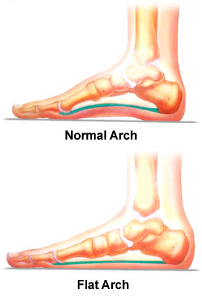
القدم المفلطحة FLATFOOT PES PLANUS


القدم المفلطحة flexable flatfoot
Pes planovalgus))
يعتبر هذا النوع من اكثر الحالات شيوعا عند الاطفال وهي تتكون من
1/ decrease in the medial longitudinal arch
2/ valgus hindfoot
3/ forefoot abduction with weightbearing
incidence  :
 unknown in pediatric population
 20% to 25% in adults
Pathoanatomy:
generalized ligamentous laxity ليونة عامة في الاربطة
25% are associated with gastrocnemius-soleus contracture وفي 25% من الحالات تكون مرتبطة مع عضلات بطة الساق
Prognosis:
غالبا ما تتحسن مع الوقت
Classification:
Hypermobile flexible pes planovalgus هذا النوع الاكثر شيوعا
عائلي familial
غالبا ما يكون في كلتا القدمين ويكون مرتبط بليونة عامة في الاربطة
associated with an accessory navicular
النوع الثاني : Flexible pes planovalgus with a tight heel cord
النوع الثالث: Rigid flatfoot & tarsal coalition (least common)
النوع الثالث هو نوع معقد ولا يتحسن مع الوقت وهو يعاني من محدودية الحركة في مفصل subtalar
عادة لا يشكو من اعراض او يشكو من الم خفيف في منطقة القوس في بطن القدم او حول الساق
التشخيص diagnosis:
من خلال الفحص الاكلينيكي  والاشعة
العلاج treatment:
قد يحتاج علاج تحفظي ومتابعة او علاج جراحي
 

ACHOHDROPLASIA


Achondroplasia قصر قامة /قصر الاطراف

is a  disproportionate short-limb  dwarfism

الظهور 3 حالات لكل 100000

عند الاطفال حديثي الولادة INFANTS يعانو من HYPOTONIA ويعانو ايضا من تسديد في القناة التنفسية العلوية وضغط علي النخاع الشوكي مما يسبب حالات الموت

يتراوح الطول عند الذكور المصابين ما بين 131+5.6سم وعند الاناث 124+5.9سم

الفحص او الشكل العام لهذه الحالات تكون كالتالي :

1/ Exaggerated lumbar lordosis انحناء كبير للجزء القطني من العمود الفقري


2/ , limitation of elbow extension and rotation تقيد في حركة مفصل الكوع

3/ , genu varumتقوس في الساقين

4/ , hyperextension of the knees and most other joints is commonشد خلفي في معظم مفاصل الجسم.

الجمعة، 20 مايو 2016

جراحة العظام والمفاصل .كسر الفخذ femor fracture

orthopedic and  traumatology 


جراحة العظام والمفاصل

fracture femorكسر الفخد  

 الكسر هو انفصال في المد الطبيعي للعظم 
braek of the bone continuety 

وهذا لكسر هو من الكسور التي تتردد باستمرار علي المستشفيات 
وتختلف طرق علاجه حسب عمر المريض ومكان الكسر ونوعية الكسر 
فمثلا عند الاطفال نستخدم علاج الكسر باستخدام مادة الجبس علي شكل بنطلون 

وفي الفئات العمرية الاكبر نستخدم طرق العلاج بالصفائح والمساميروالاسياخ البلاتينية 

femour fracture one of most typeof fractures 

and the managment is defferance according to age of patients and type or localisation of fracture 

in children most typeof managment is a hip spica 

in the other age group we are use alot of methods as 
plating and scrwes

interlocking nail .dynamic hip screw,austein moore replacement